Reseña o resumen
A convenient and authoritative quick-reference guide to help you get the most from radiography of dogs and cats.
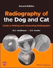
In the newly revised second edition of Radiography of the Dog and Cat: Guide to Making and Interpreting Radiographs, the authors deliver a thorough update to a celebrated reference manual for all veterinary personnel, student to specialist, involved with canine and feline radiography. The book takes a straightforward approach to the fundamentals of radiography and provides easy-to-follow explanations of key points and concepts. Hundreds of new images have been added covering normal radiographic anatomy and numerous diseases and disorders.
This latest edition includes access to a companion website with review questions and answers. Readers of the book will also find:
An expanded positioning guide along with images of properly positioned radiographs.
Numerous examples of radiographic artifacts with explanations of their causes and remedies.
Detailed explanations of many contrast radiography procedures, including indications, contraindications, and common pitfalls.
Comprehensive treatments of Musculoskeletal, Thoracic, and Abdominal body parts, including both normal and abnormal radiographic appearances and variations in body types.
Perfect for veterinary practitioners and students, the second edition of Radiography of the Dog and Cat: Guide to Making and Interpreting Radiographs is also a valuable handbook for veterinary technical staff seeking a one-stop reference for dog and cat radiography.
M. C. Muhlbauer, DVM, MS, DACVR is a veterinary radiologist with over 30 years of experience in teaching and performing diagnostic imaging. He is owner and president of Veterinary Imaging Specialists in Venice, Florida, USA.
S. K. Kneller, DVM, MS, DACVR retired as Associate Professor Emeritus from the University of Illinois College of Veterinary Medicine in Urbana in 2007 after over 32 years and since then has served as Locum Tenens at eight different North American veterinary colleges and as Adjunct Instructor at Midwestern University College of Veterinary Medicine, Glendale, Arizona, USA.
About the Companion Website
Acknowledgments
Introduction
Chapter 1 X-rays
Properties of x-rays
X-ray production
X-ray machine
Image receptors
Geometry of the x-ray beam
X-ray interactions with matter
Radiographic density
Opacity
Radiographic contrast
Radiographic detail
Technique chart
Radiograph storage and distribution
Radiation Safety
Chapter 2 Radiographs
Orthogonal views
Procedure for making radiographs
Nomenclature
Positioning Guide
Thoracic
Abdomen
Musculoskeletal
Artifacts
Contrast radiography
Reading radiographs
Chapter 3 Thorax
Making thoracic radiographs
Patient factors
Thoracic wall
Diaphragm
Pleural and pleural space
Mediastinum
Esophagus
Heart and major vessels
Trachea
Lungs
Chapter 4 Abdomen
Making abdominal radiographs
Patient factors
Abdominal cavity
Liver
Spleen
Pancreas
GI tract
Stomach
Small intestine
Large intestine
Urogenital tract
Kidneys and ureters
Urinary bladder
Urethra
Male genital system
Female genital system
Adrenal glands
Chapter 5 Musculoskeletal
Making musculoskeletal radiographs
Soft tissues
Orthopedic anatomy
Bone response to disease or injury
Fractures
Generalized musculoskeletal diseases
Benign conditions of bone
Congenital and developmental abnormalities
Joints
Appendicular skeleton
Shoulder
Elbow
Carpus
Digits
Pelvis
Stifle
Tarsus
Axial skeleton
Vertebral column
Head and neck
Glossary
Index